Fetal Scans

Services
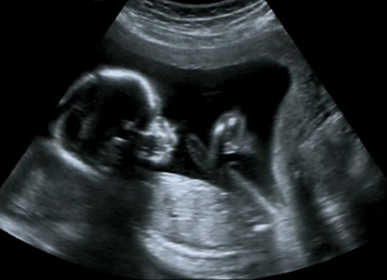
Fetal ultrasound is a test used during pregnancy that creates an image of the fetus in the mother’s uterus, or womb. During a fetal ultrasound, various parts of the baby, such as the heart, head, and spine, are identified and measured. The testing may be performed either through the mother’s abdomen (transabdominal) or vaginal canal (transvaginal). Fetal ultrasound provides a safe way to evaluate the health of an unborn baby.
There are several types of fetal ultrasound, each with specific advantages in certain situations. A Doppler ultrasound, for example, helps to study the movement of blood through the umbilical cord between the fetus and placenta. Three-dimensional ultrasound provides a life-like image of an unborn baby. The standard, 2-dimensional ultrasound is the primary focus of this discussion, but the basic concepts for standard fetal ultrasound also apply to the other types.
Keeping the Parents Informed

3D & 4D Scans

Absent Nasal Bone

Aminocentesis

First Trimester Scan

Chorionic Villus Sampling

Chorionic Villus Sampling

Pre-Eclampsia

Short Cervix

Fetal Anemia

Fetal Reduction

Gastroschisis

Genetics

Down's Syndrome

Multiple Pregnancy

Neural Tube Defects

Omphalocele

SELECTIVE FETAL GROWTH RESTRICTION (sFGR)

NIPT- NON INVASIVE PRENATAL TEST

Twin To Twin Transfusion Syndrome (TTTS)

Intra-Uterine Blood Transfusion

